Milia: Causes, Look-Alikes & When to Get Them Checked
What milia are, what causes them, how to tell them apart from look-alike lesions, whether the NHS removes them, and what treatment costs in Bedford.
Milia are small, firm, pearly-white bumps caused by keratin sealed just beneath the skin, not a type of spot, and not something a pore lets you squeeze out. Most newborn milia clear by themselves within weeks; milia that appear later in life can persist for months or years, which is why they are usually cleared privately rather than left.
What are milia?
Milia are small, benign, keratin-filled cysts that sit just beneath the surface of the skin. StatPearls describes them as “benign and transient subepidermal keratin cysts”, usually presenting as tiny, firm, white papules on the face, though they can also appear on the upper trunk, limbs and genital area. DermNet NZ puts it more plainly: milia are “harmless cysts [that] present as tiny pearly-white bumps just under the surface of the skin.”
They are extremely common in newborns (StatPearls records that “primary congenital milia occur in up to 40% to 50% of healthy full-term neonates”), but they also occur in older children and adults, most often around the eyelids, cheeks and forehead.
Dermatologists split milia into two broad groups. Primary milia arise spontaneously on otherwise healthy skin, thought to originate from the sebaceous collar of vellus (fine, soft) hairs. Secondary milia form where skin has been damaged or occluded (after trauma, blistering skin conditions, some cosmetic procedures, certain medications, or with heavy, occlusive skincare) and per StatPearls “are believed to derive from eccrine ducts more commonly than from overlying epidermis, hair follicles, or sebaceous ducts.”
Why milia look pearly white
A milium is a tiny keratin-filled cyst just below the skin surface. Around the eyelids and cheeks it often appears as a firm pearly-white bump.

- Surface view
Small white or pearly bumps beneath intact skin.
- Skin-layer view
A simplified pocket of keratin immediately below the epidermis.
What causes milia?
Primary milia are thought to develop from the lower part of a vellus hair follicle, sealing a small pocket of keratin (the structural protein that makes up the outer layer of skin) just beneath the surface.
Secondary milia form after something disrupts the skin’s normal shedding: sunburn, blistering skin disease, dermabrasion or laser resurfacing, certain topical medications, or ordinary skin trauma can all trigger them.
A small number of rare inherited conditions (including Brooke–Spiegler syndrome, Bazex–Dupré–Christol syndrome and Rombo syndrome) are associated with unusually widespread or persistent milia, per the standard classification review (Berk & Bayliss, JAAD, 2008). These are uncommon, but worth knowing about if milia are extensive, unusual in distribution, or run in the family alongside other skin features.
Is milia dangerous? When to get it checked
Ordinary milia are not dangerous. None of the sources reviewed for this page (a peer-reviewed clinical classification, two clinical reference resources, and an NHS commissioning policy) flag milia as a marker of any internal disease or malignancy.
The real safety point is diagnostic, not oncological: several other lesions can look like milia, and they are managed differently.
- Sebaceous hyperplasia: enlarged oil glands that form similar white-to-yellow bumps, usually around the nose and upper lip, but with a different (glandular) origin and often a visible central dell.
- Syringomas and xanthelasma: both common around the eyelids, and both distinct conditions with their own causes and treatment considerations.
- Comedonal acne, flat warts and calcinosis cutis: other look-alikes worth ruling out before treatment.
- Milia en plaque: a less common variant where milia coalesce into a plaque rather than staying as discrete small bumps, which has genetic associations worth a doctor’s opinion if it appears.
See a doctor rather than book cosmetic extraction if a bump is atypical, rapidly changing, forming a plaque rather than a discrete cyst, or you are simply not sure what it is. A firm, discrete, pearly-white, non-inflamed bump that has looked the same for weeks or months is the classic, low-concern picture, but that judgement is exactly what a proper assessment is for.
Can you get milia removed on the NHS?
Not for cosmetic reasons.
An NHS Integrated Care Board commissioning policy (Gloucestershire ICB, “Removal of Benign Skin Lesions”, v1.2, adopted 2024) names milia specifically among the benign lesions for which it will not fund a hospital referral. The policy states: “Gloucestershire ICB will not routinely fund referral to secondary care for the treatment of benign skin lesions, including all the following conditions: Warts, Milia, Sebaceous hyperplasia, Seborrhoeic keratoses, Spider Naevi/cherry angiomas…” Funding is only considered where a lesion is genuinely obstructing an orifice, causing pain from unavoidable pressure, bleeding repeatedly from unavoidable trauma, or documented as recurrently infected: none of which milia typically cause.
Two things are worth being honest about here, because they are unusual gaps for such a common condition. There is no dedicated NHS.uk patient page on milia (a direct check returns a 404), and no British Association of Dermatologists patient leaflet on milia could be found either, despite a targeted search of their leaflet library. That is not a case of failing to find the guidance: the guidance does not appear to exist in either place. So if you are looking for NHS-endorsed information specifically about milia, there genuinely isn’t a dedicated official page to point you to; this page is written from peer-reviewed clinical references (StatPearls, a JAAD classification review, DermNet NZ) instead.
There is no single fixed price, because it depends on the number of milia, where they are and which method is used. Rejuvena’s own milia removal page states plainly: “Cost depends on the number, location and method, so your price is confirmed at your consultation with no obligation to proceed.”
For context, Rejuvena’s general laser skin lesion removal page (which lists milia among the lesions it treats) states pricing “From £150, priced by lesion size and number, with a cosmetic skin lesion assessment at £50.” That figure covers the wider lesion-removal service rather than milia specifically, so treat it as a starting reference point rather than a milia quote.
How milia removal works at Rejuvena
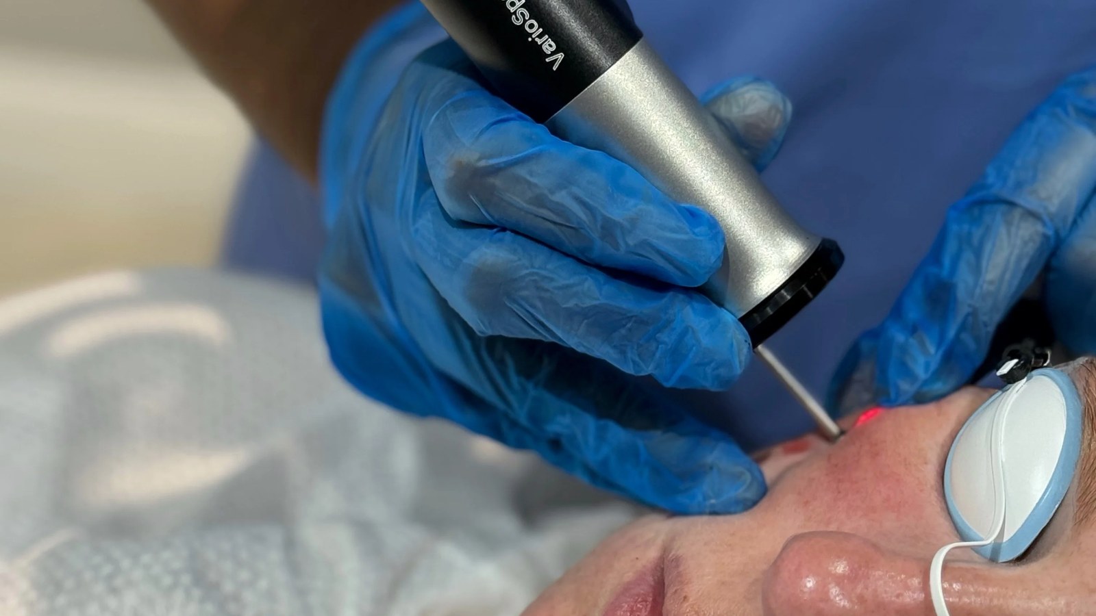
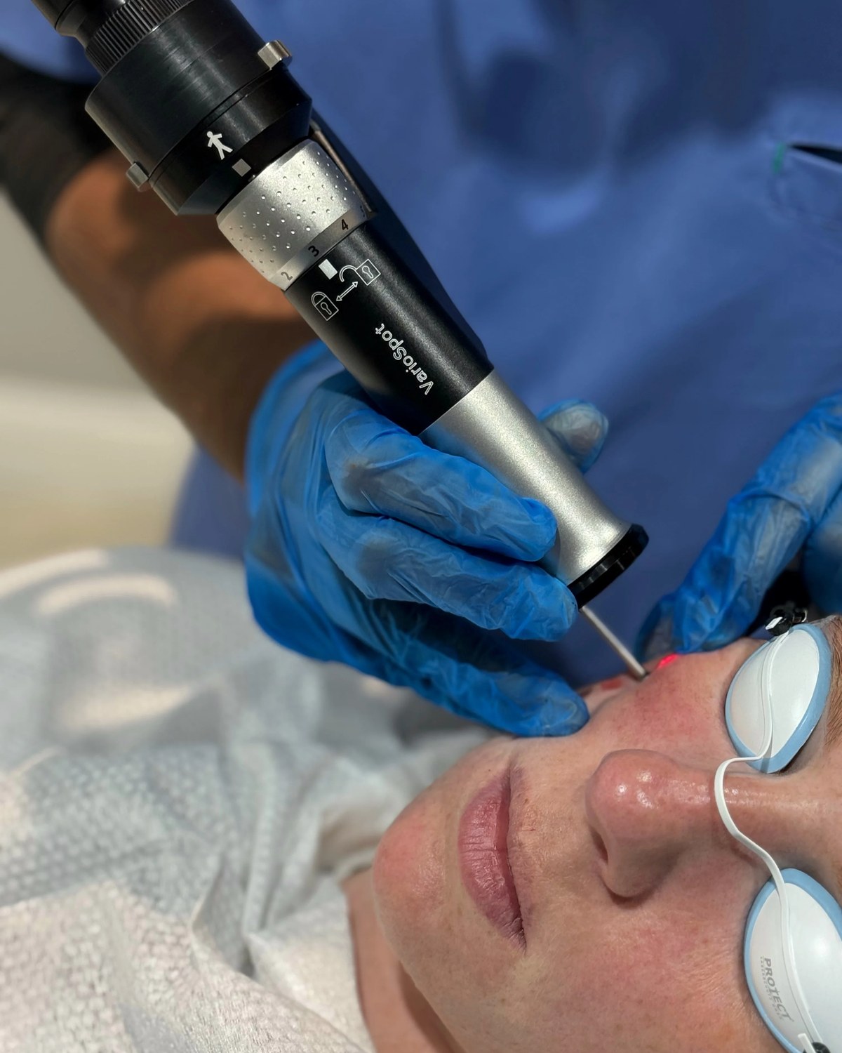
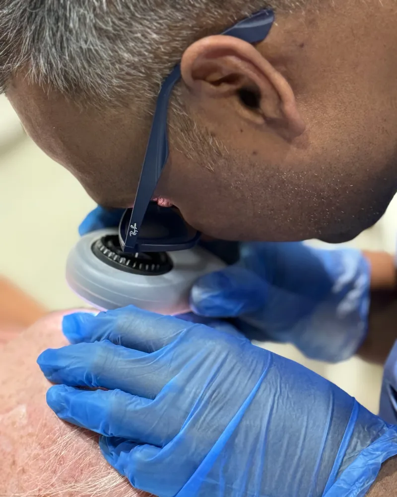
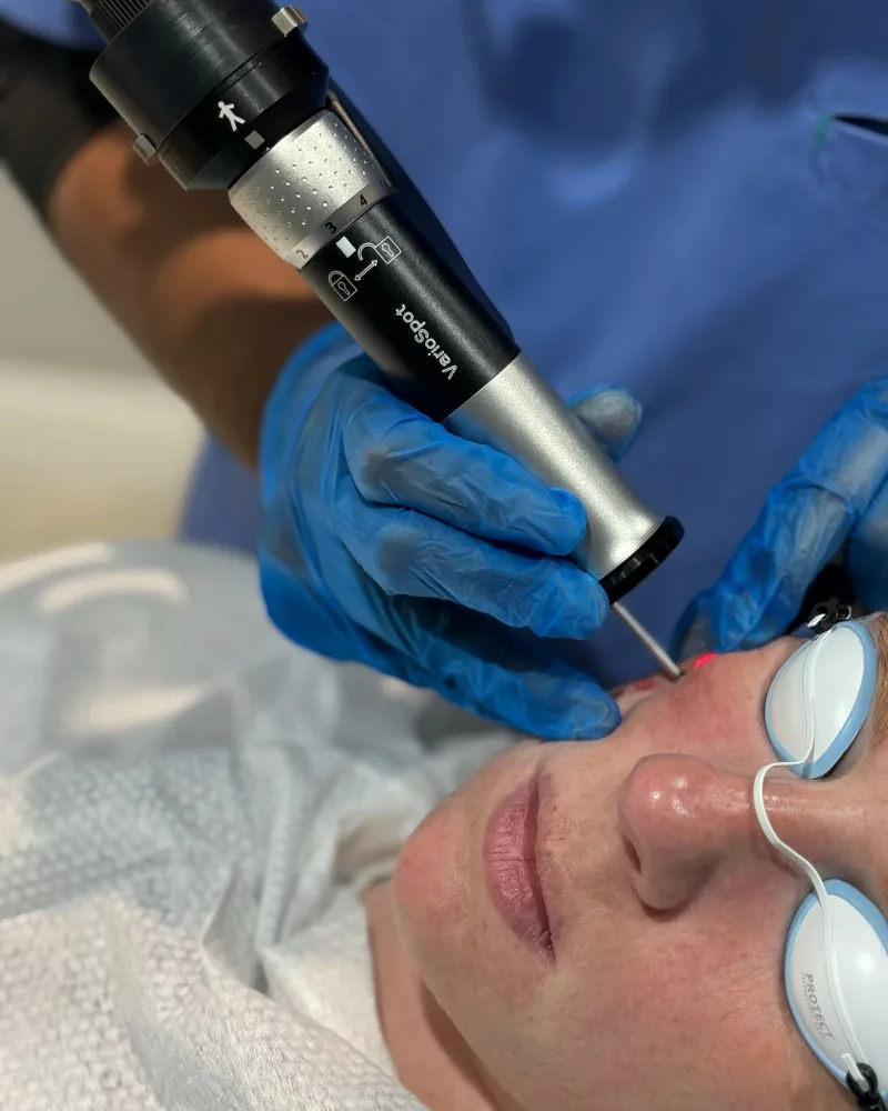
Every bump is examined by Dr Nick first, to confirm it is genuinely a milium rather than a look-alike. Small, superficial milia and clusters are typically treated by sterile de-roofing and extraction: a fine lancet opens the cyst and the keratin core is gently lifted out. Deeper, persistent or awkwardly placed milia can instead be treated with the Dermablate Cool Laser™ (Er:YAG, 2940nm), which ablates a controlled micro-layer with minimal heat spread: precise enough for use close to the eye.
Why choose Rejuvena for milia assessment in Bedford?
Diagnosis first
Every bump is examined before treatment, to rule out sebaceous hyperplasia, syringoma and other look-alikes.
Diploma in Dermatology
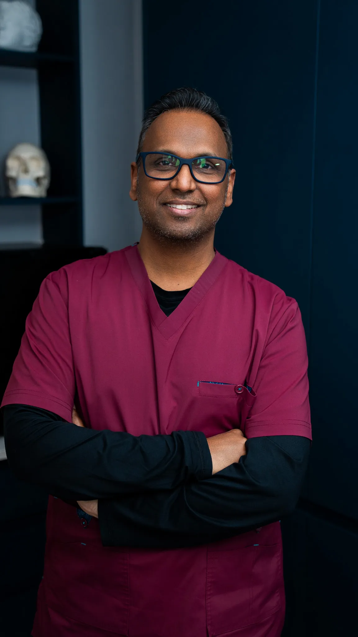
Assessed by Dr Nick, a GMC-registered doctor, not a beauty therapist or nurse.
Precise near the eye
The Cool Laser™ Er:YAG ablates in controlled micro-layers with minimal heat spread, for delicate placements.
Local & welcoming
Based at 52 Thor Drive, Bedford, we care for patients from Kempston, Biddenham, Bromham and across Bedfordshire.
Verified Google reviews
What our patients say
5.0from 161 Google reviews
I recently had a lesion removed from my cheek. The pigmented lesion had bothered me for years. I’m glad I finally decided to do something about it. I was quite nervous on the day, but Dr. Nick and Lana were so careful and reassuring — they really put me at ease. The procedure was quick and comfortable, and I feel so much better for having it done. I did a lot of research before I chose this clinic for my treatment. The clinic’s medical dermatology focus made me feel confident I would be in the right hands. I’m really grateful for the care I received and I would definitely recommend this clinic.
Rejuvena repliedThank you so much for your skin review Dupinder 🙏
Read this review on Google (opens in a new tab)I had lots of skin lesions on both hands and Dr Nick and Lana have been great treating me.I will definitely be returning for any future treatments and highly recommend seeing Dr Nick.Great service and facilities thanks again.
Rejuvena repliedAww! Thank you Gavin for your kind review. It was a great pleasure having you in our clinic and we look forward to seeing you in the future too. 😊🙏
Read this review on Google (opens in a new tab)Thank you so much for your help, I cannot fault Lana and Dr Nick at all, the services offered were excellent and they are clearly both very caring, professional and passionate in what they do. The venue is perfect, very clean, calming and peaceful. I would highly recommend to friends and family.
Rejuvena repliedThank you for your kind words Clara. Glad we could help with your skin concerns. 😊🙏
Read this review on Google (opens in a new tab)Lana is very knowledgeable and professional and makes you feel like you are in safe hands. I am happy I chose to have my treatment at Rejuvena and would highly recommend the clinic.
Rejuvena repliedThank you very much Bobbie 🤗🙏
Read this review on Google (opens in a new tab)Very good and reassuring place to book your next or target any problem. This is where you want to be.
Rejuvena repliedAww! Thank you Peter. That means a lot to us 😊🙏
Read this review on Google (opens in a new tab)Lana is a complete professional and wonderful at what she does! I always feel in safe hands knowing her attention to detail is spot on and she genuinely cares about doing a great job for her clients.
Rejuvena repliedThank you very much Daisy for your kind words. ☺️🙏
Read this review on Google (opens in a new tab)Every review above is published on Google. Tap any card to read it there.
Milia FAQs in Bedford
How much does milia removal cost in the UK?
Do the NHS remove milia?
Will a GP remove milia?
Why won’t milia just pop like a spot?
Book your milia assessment in Bedford
Speak with Dr Nick’s team at Rejuvena Bedford for a doctor-led milia assessment. Book online or call us.