Cherry Angiomas: Causes, Safety & When to Get Checked
What cherry angiomas are, what causes them, the eruptive-pattern melanoma association, whether the NHS removes them, and what treatment costs in Bedford.
A cherry angioma (also called a Campbell de Morgan spot) is a common, harmless overgrowth of small blood vessels that shows as a bright-red spot, usually on the chest or trunk. Most people simply have one or a few. A sudden crop of many new spots in a short time is worth mentioning to a doctor.
What are cherry angiomas?
A cherry angioma is a benign, acquired vascular skin lesion: the Primary Care Dermatology Society (PCDS) defines it as “a benign acquired vascular proliferation of the skin”, with the term “cherry angioma” used for lesions with “a cherry-like (or blackberry-like) appearance.” An NHS patient leaflet (Wrightington, Wigan and Leigh NHS Foundation Trust) describes them more simply: “little reddish spots… caused by small areas of blood vessel overgrowth.”
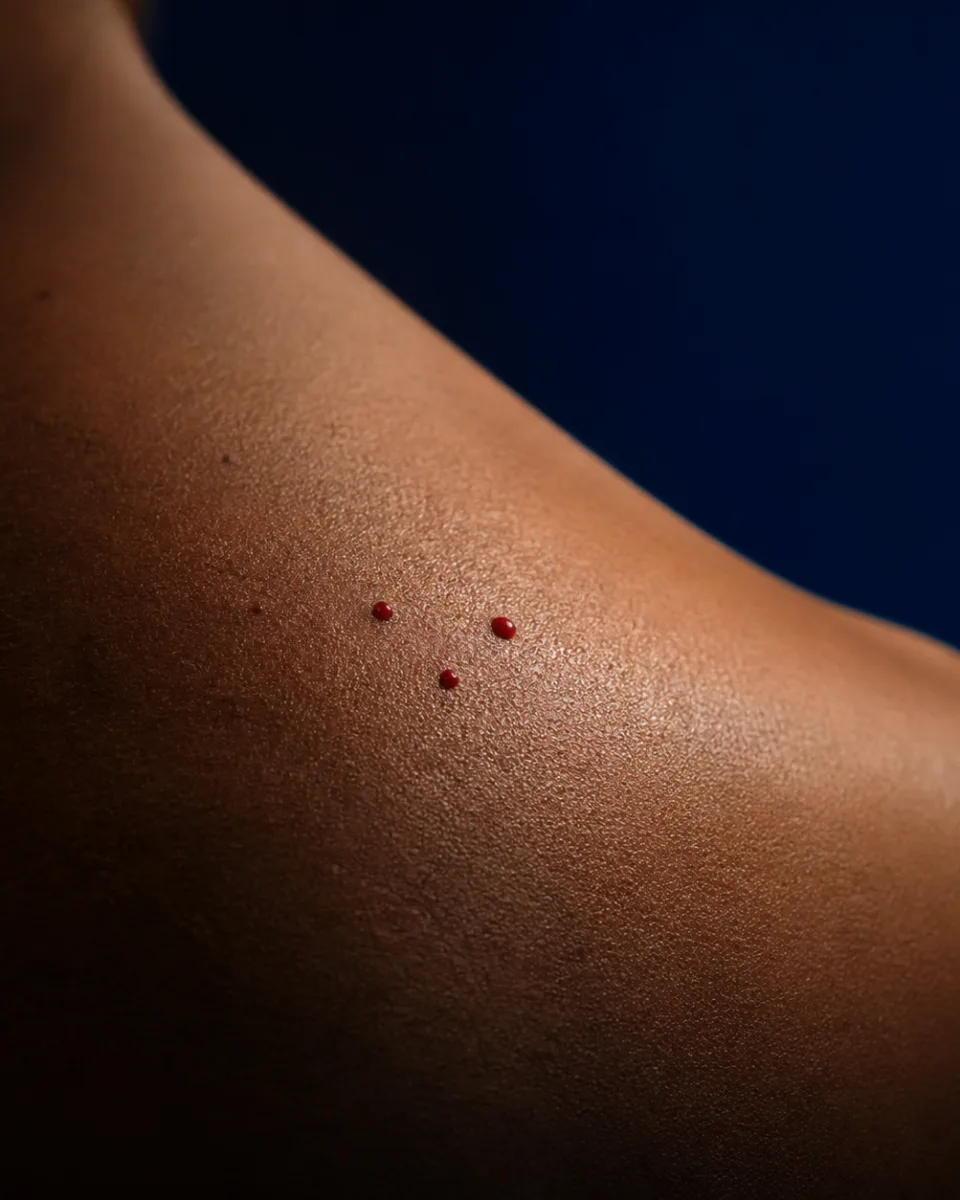
They are small, discrete, cherry-red to purplish dome-shaped spots, usually 0.5–6mm across, most often on the chest, trunk and upper limbs, though they can appear anywhere. They become steadily more common with age, and it is normal to have several. They can occasionally bleed if caught or knocked, or suddenly turn black if a lesion thromboses (“surface bleeding, or a bleed into the surrounding skin, can occur if traumatised”, per PCDS): both of which sound alarming but are recognised, benign features of the lesion itself.
Why a cherry angioma looks red
A cherry angioma is a small vascular papule. Its red, blue or purple colour comes from a collection of superficial capillaries.

- Surface view
A small, smooth, firm red-to-purple papule.
- Skin-layer view
A simplified cluster of dilated capillaries in the superficial dermis.
What causes cherry angiomas?
They form from proliferating, dilated capillaries and small veins in the upper layer of skin (the papillary dermis). The precise trigger for this vessel overgrowth is not established in the sources reviewed for this page. What is well documented is that numbers increase with age, and they are sometimes linked to hormonal changes such as pregnancy, and to a tendency that runs in families. Unlike sun spots, they are not related to sun exposure: the NHS Wigan leaflet is explicit: “They are not pre-cancerous and are not related to sun exposure.”
Is a cherry angioma dangerous? When to get it checked
An ordinary cherry angioma (a single spot, or a few that have built up gradually over years) is not dangerous, is not pre-cancerous, and is not related to sun exposure, per the NHS Wigan leaflet above.
There is one genuine pattern worth being aware of, checked directly against the published research for this page rather than taken on trust. A peer-reviewed cross-sectional study (Borghi et al., Dermatology, 2021, n=1,190) found that an eruptive pattern of cherry angiomas (defined in that study as more than 10 lesions) was significantly associated with malignant melanoma, particularly in people under 70, with a strength of association the authors describe as “similar” to the well-established link between atypical moles and melanoma. This is an association from an observational study in a dermatology-oncology setting, not proof that eruptive cherry angiomas cause or diagnose melanoma, but it is real, published, peer-reviewed evidence.
Crucially, this applies to a sudden crop of many new spots appearing over a short period, not to the ordinary, gradual accumulation of one or a few spots that most people have. A more recent case series reviewing the wider literature (Campo et al., Cureus, 2025) reached a reassuring conclusion for otherwise healthy people: “reassurance and discussion early on with patients will help reduce patient distress, unnecessary follow-ups, and waste of resources”: the point is patient education and a routine skin check, not alarm or automatic cancer screening.
Get a spot checked rather than book cosmetic removal if it is changing, growing, bleeding without being knocked, ulcerating, turning brown or an irregular colour, or has visible vessels running through it in an unusual pattern: PCDS flags these as signs the lesion might not be an ordinary cherry angioma at all, with melanoma one of the differentials to rule out. And if you notice many new spots appearing over weeks rather than years, that eruptive pattern is worth a GP conversation, particularly if you are under 70.
Can you get a cherry angioma removed on the NHS?
Not for cosmetic reasons. PCDS is direct: “Treatment is not normally required. Lesions that catch/bleed can be removed by curettage and sent for histology” in other words, NHS removal is reserved for spots causing genuine physical problems, and even then the tissue is sent for laboratory analysis as a precaution.
An NHS Integrated Care Board commissioning policy (Gloucestershire ICB) lists “Spider Naevi/cherry angiomas” explicitly among the benign lesions it will not routinely fund a hospital referral for. As with other benign lesions, funding is only considered for genuine functional problems (obstruction, pain from unavoidable pressure, repeated bleeding from unavoidable trauma, or documented recurrent infection) not for appearance alone.
There is no single fixed price. Rejuvena’s cherry angioma removal page states: “the price is confirmed at your consultation once Dr Nick has assessed the lesion and agreed a plan with you,” because the method, the number of spots and the number of sessions needed all vary from person to person.
For context, the clinic’s general laser skin lesion removal page states pricing “From £150, priced by lesion size and number, with a cosmetic skin lesion assessment at £50”: a starting reference point for the wider lesion-removal service, not a cherry-angioma-specific quote.
How cherry angioma removal works at Rejuvena
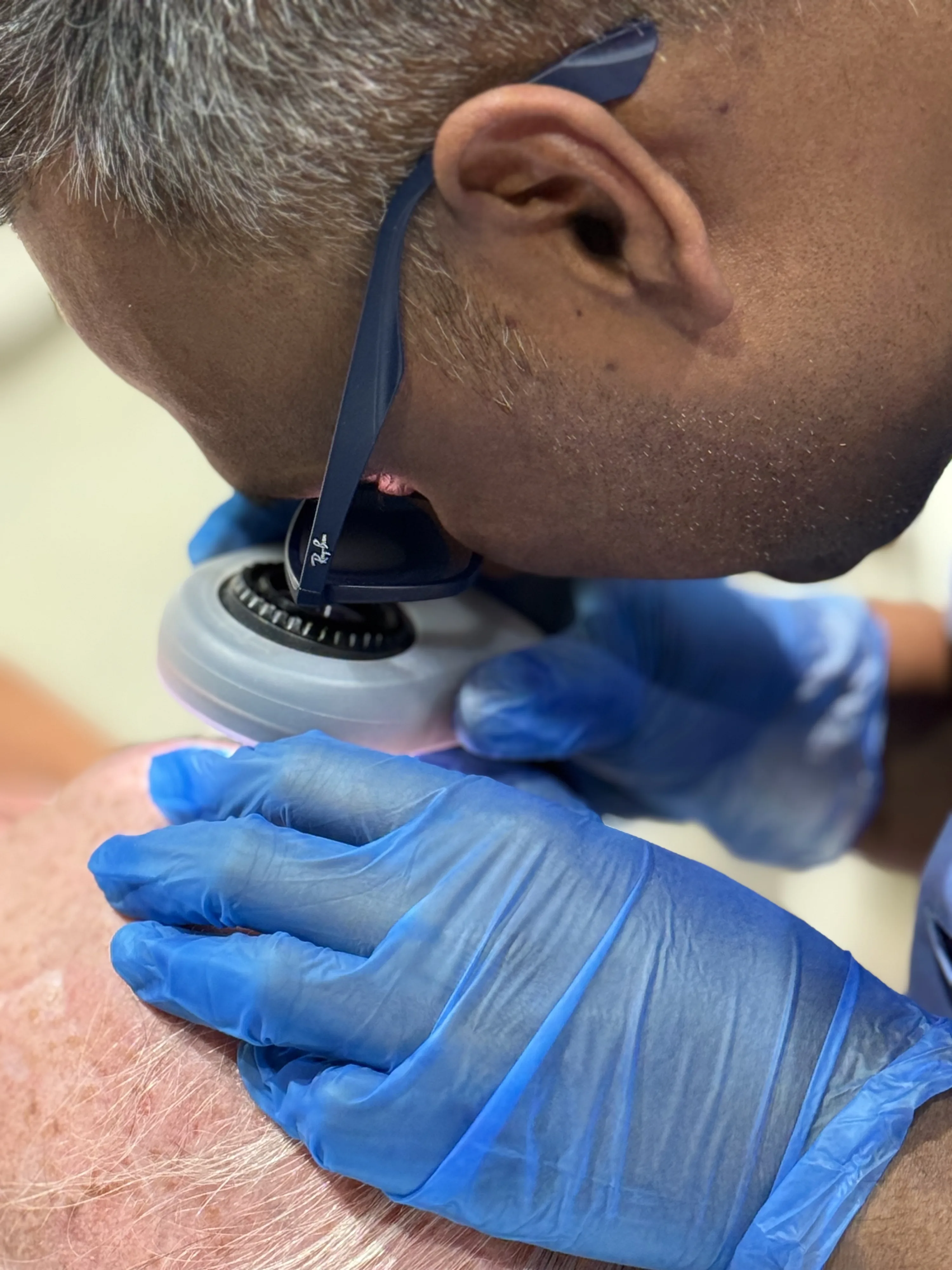
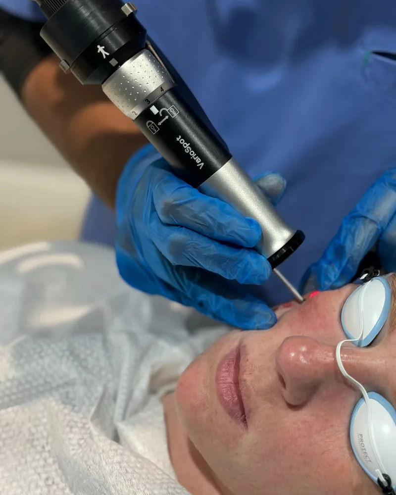
Every lesion is assessed by Dr Nick first (with dermoscopy where appropriate) to confirm it is a benign cherry angioma rather than a spider naevus, angiokeratoma, pyogenic granuloma or anything needing referral. Smaller spots, or several spots at once, are typically treated with IPL/vascular light, which targets the blood inside the vessel and seals it. A single raised lesion is often better suited to a targeted laser/lesion-removal technique. The right method for your spot is confirmed at consultation.
Why choose Rejuvena for cherry angioma assessment in Bedford?
Diagnosis first
Every lesion is assessed, with dermoscopy where appropriate, before any spot is treated cosmetically.
Diploma in Dermatology
Assessed by Dr Nick, a GMC-registered doctor, not a beauty therapist or nurse.
Referred, not ignored
An eruptive pattern of new spots is flagged for a GP conversation, not simply lasered away.
Local & welcoming
Based at 52 Thor Drive, Bedford, we care for patients from Kempston, Biddenham, Bromham and across Bedfordshire.
Verified Google reviews
What our patients say
5.0from 161 Google reviews
Lana and Dr Nick are both friendly and professional. I had treatment for thread vein removal and starting to see good results already! Thank you :)
Rejuvena repliedThank you very much for your kind words Alison. Your thread veins are improving great! Kind regards 🤗
Read this review on Google (opens in a new tab)I recently had a lesion removed from my cheek. The pigmented lesion had bothered me for years. I’m glad I finally decided to do something about it. I was quite nervous on the day, but Dr. Nick and Lana were so careful and reassuring — they really put me at ease. The procedure was quick and comfortable, and I feel so much better for having it done. I did a lot of research before I chose this clinic for my treatment. The clinic’s medical dermatology focus made me feel confident I would be in the right hands. I’m really grateful for the care I received and I would definitely recommend this clinic.
Rejuvena repliedThank you so much for your skin review Dupinder 🙏
Read this review on Google (opens in a new tab)I had lots of skin lesions on both hands and Dr Nick and Lana have been great treating me.I will definitely be returning for any future treatments and highly recommend seeing Dr Nick.Great service and facilities thanks again.
Rejuvena repliedAww! Thank you Gavin for your kind review. It was a great pleasure having you in our clinic and we look forward to seeing you in the future too. 😊🙏
Read this review on Google (opens in a new tab)Where to begin? Easy online booking, friendly team and professional pre-treatment consultations. Dr Nick’s medical background offers the expertise and reassurance that’s often missed in beauty salons. Lots of valuable advice as I explored thread vein treatments, options, budget, and desired results. In addition to the treatment itself, I’ve welcomed the superb after-care and support. I’ll absolutely be returning and already have my eye on their cool later facial. Thank you team Rejuvena!
Rejuvena repliedThank you very much Gemma, for your kind review. We are so glad to hear you liked our services. We are very grateful. ☺️🙏
Read this review on Google (opens in a new tab)Lana is very knowledgeable and professional and makes you feel like you are in safe hands. I am happy I chose to have my treatment at Rejuvena and would highly recommend the clinic.
Rejuvena repliedThank you very much Bobbie 🤗🙏
Read this review on Google (opens in a new tab)Very good and reassuring place to book your next or target any problem. This is where you want to be.
Rejuvena repliedAww! Thank you Peter. That means a lot to us 😊🙏
Read this review on Google (opens in a new tab)Every review above is published on Google. Tap any card to read it there.
Cherry Angiomas FAQs in Bedford
How much does it cost to have one cherry angioma removed?
How much does cherry angioma removal cost in the UK?
Is it worth removing cherry angiomas?
Where can I get cherry angiomas removed in the UK?
Can a sudden crop of new cherry angiomas mean something serious?
Book your cherry angioma assessment in Bedford
Speak with Dr Nick’s team at Rejuvena Bedford for a doctor-led cherry angioma assessment. Book online or call us.