Doctor-led cherry angioma removal in Bedford
Assessment-first removal of confirmed benign cherry angiomas, also called red spots or Campbell de Morgan spots. Dr Nick checks the lesion before recommending precise treatment and explains healing, recurrence and when referral is safer.
- Dr Nick · GMC registered
- Benign lesion assessment
- Precise treatment
- Bedford clinic

A cherry angioma is almost always harmless — but a red spot is exactly the kind of lesion a doctor should confirm before anyone removes it. Every lesion is assessed by Dr Nick first.
What is a cherry angioma?
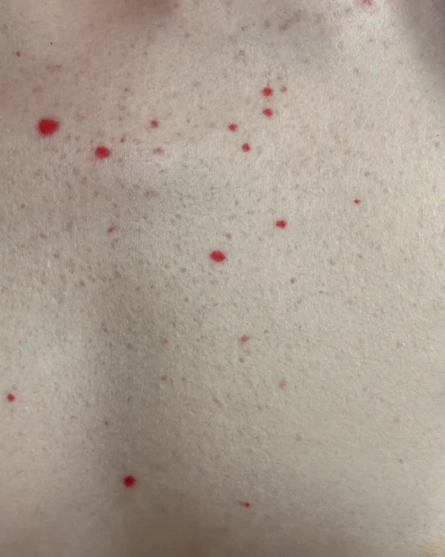
A cherry angioma — also called a Campbell de Morgan spot, red blood spot or red mole — is a common, harmless cluster of dilated small blood vessels in the skin that shows as a bright-red, blue or purple dome-shaped spot, usually 0.5–6mm across. They are a benign vascular overgrowth (proliferating capillaries in the upper layer of the skin), not skin cancer, and they most often appear on the chest, trunk, arms and sometimes the face.
They become much more common with age — around half of adults over 30 have at least one, rising to roughly three in four people by age 75 — and they can be linked to hormonal changes such as pregnancy, and to a family tendency. The exact cause is not fully understood, so we never over-claim it. Importantly, a cherry angioma is different from a thread or spider vein, which is linear or branching, and from a venous lake, which is a soft blue-purple lesion usually on the lip or ear. Telling these apart matters — which is why a proper assessment comes first.
Are cherry angiomas dangerous — and when should one be checked?
Usually harmless
A typical cherry angioma is benign and carries no cancer risk. Many people simply want one removed because it catches on clothing or jewellery, bleeds when knocked, or because they dislike how it looks.
Red flags to assess
A red or dark spot that is changing, growing, bleeding spontaneously, ulcerating, turning brown or irregular, or has vessels running through it should be examined — not simply lasered away. Benign-looking lesions can occasionally be mimicked by more serious conditions.
Assessed and referred
Dr Nick examines every lesion (with dermoscopy where appropriate) to confirm it is a benign cherry angioma. Anything atypical or of diagnostic uncertainty is assessed and referred appropriately, or sent for histology, rather than cosmetically removed.
What cherry angioma removal helps with
At Rejuvena in Bedford, each cherry angioma removal plan starts with a careful assessment to confirm the concern, suitability and realistic next steps.
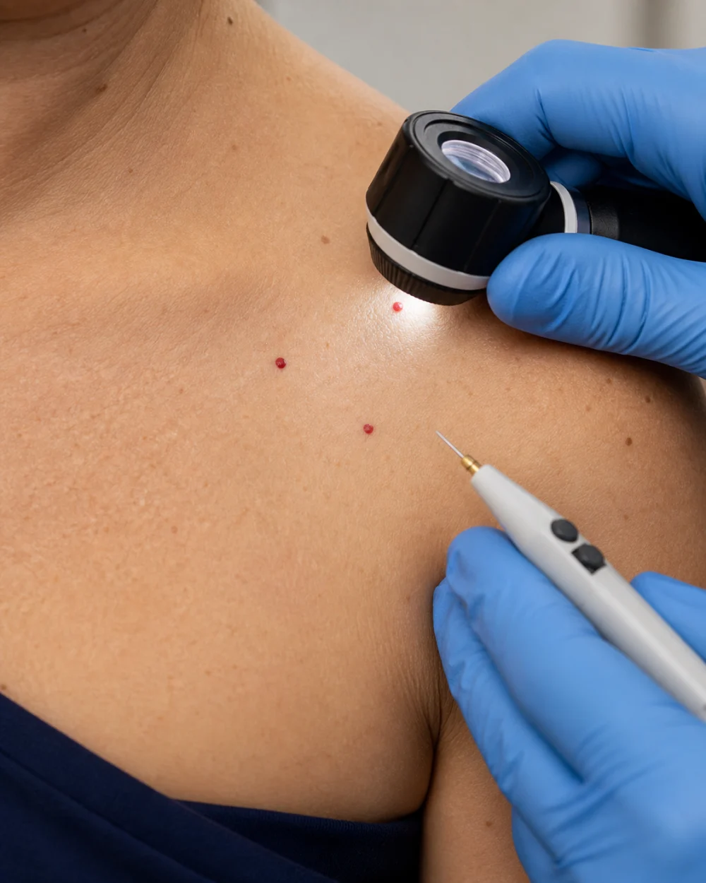
How we remove cherry angiomas at Rejuvena
There is no single “right” method — the technique is matched to your lesion at consultation. Light-based treatments target the blood inside the spot through selective photothermolysis: the light is absorbed by oxyhaemoglobin in the vessels, heating and sealing them while sparing the surrounding skin.
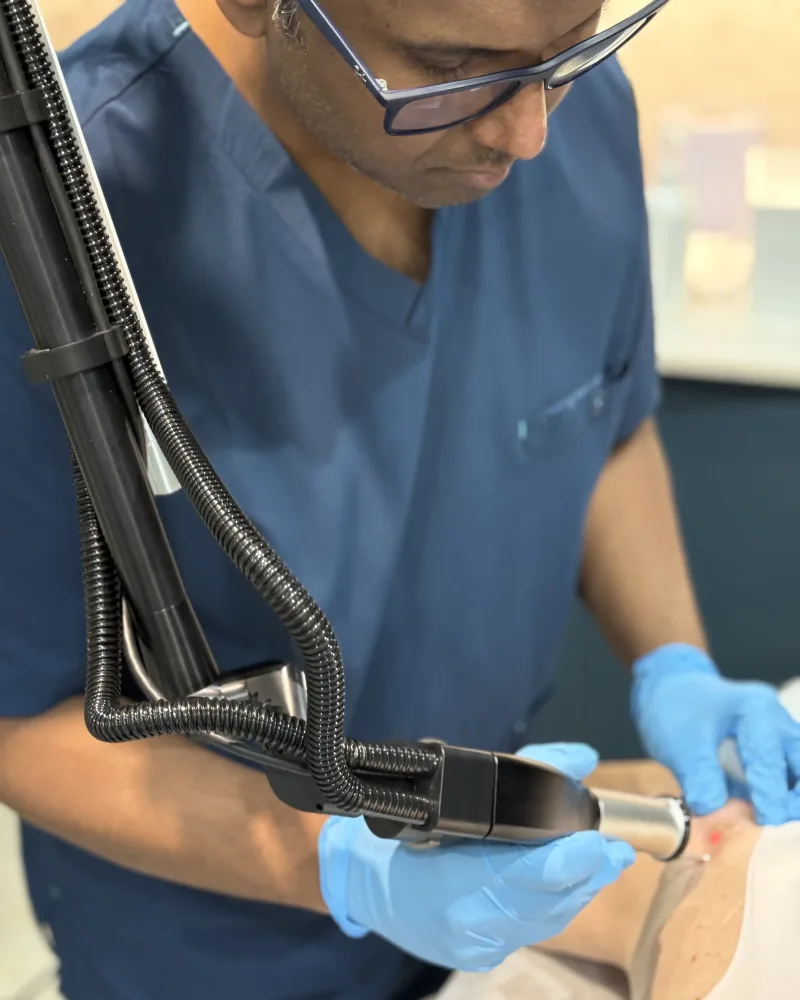
IPL & vascular light treatment
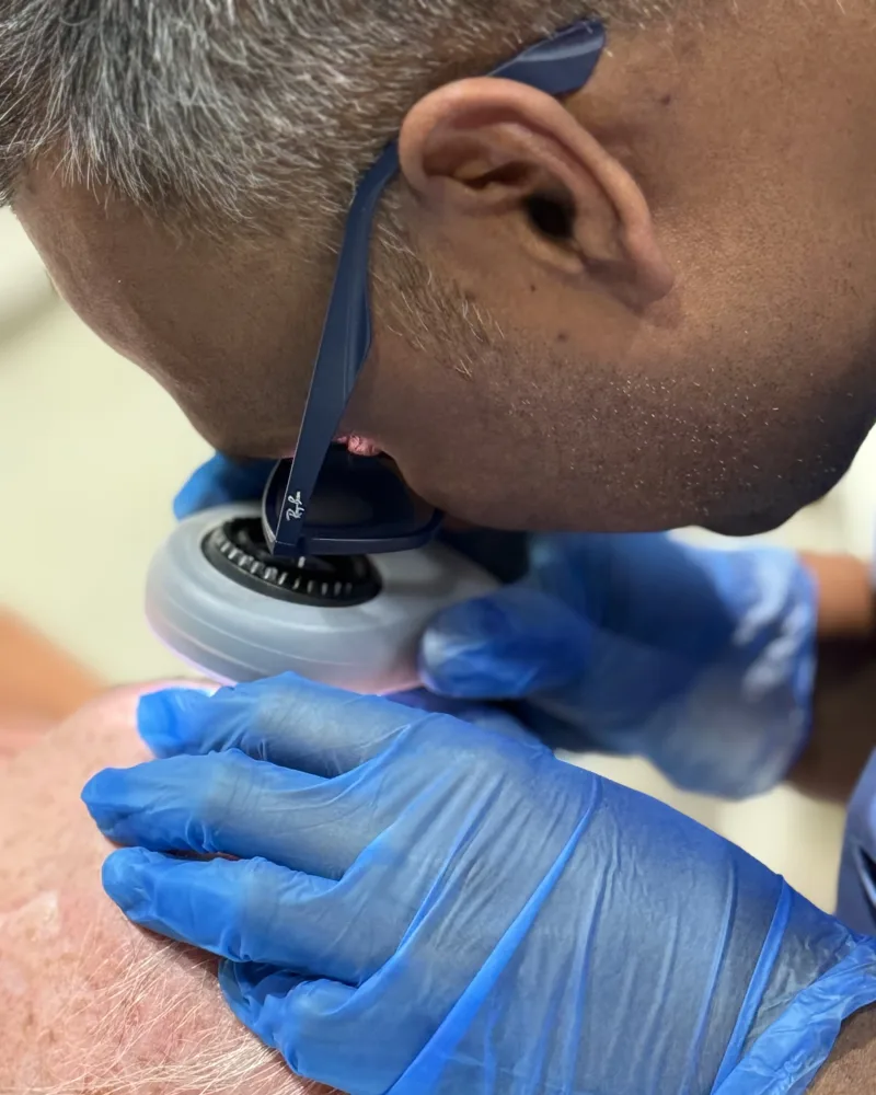
Intense pulsed light such as Lumecca IPL is well suited to smaller cherry angiomas and to people with several spots to tidy. The light energy is preferentially absorbed by the blood in the lesion, coagulating the tiny vessels so the spot fades. It tends to involve less downtime than ablative methods and may be repeated over a small number of sessions.
- Best for Smaller spots, multiple lesions
- Sessions Often 1; sometimes 2–3
- Downtime Minimal — brief redness
- How it works Light sealed onto the vessels
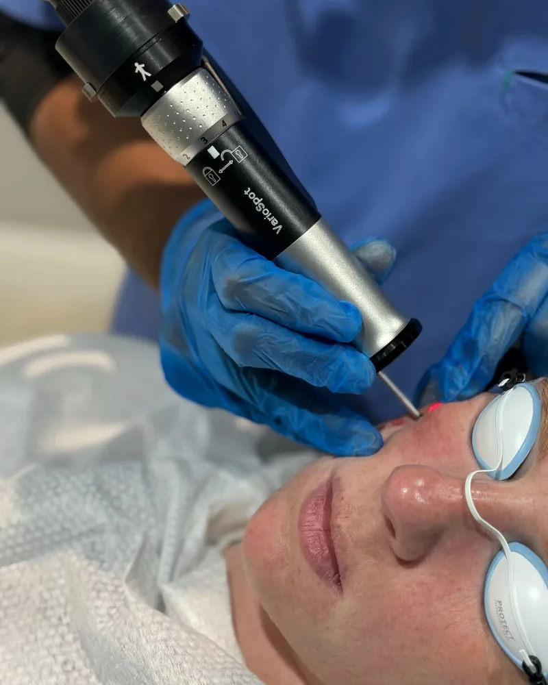
Laser / lesion-removal method
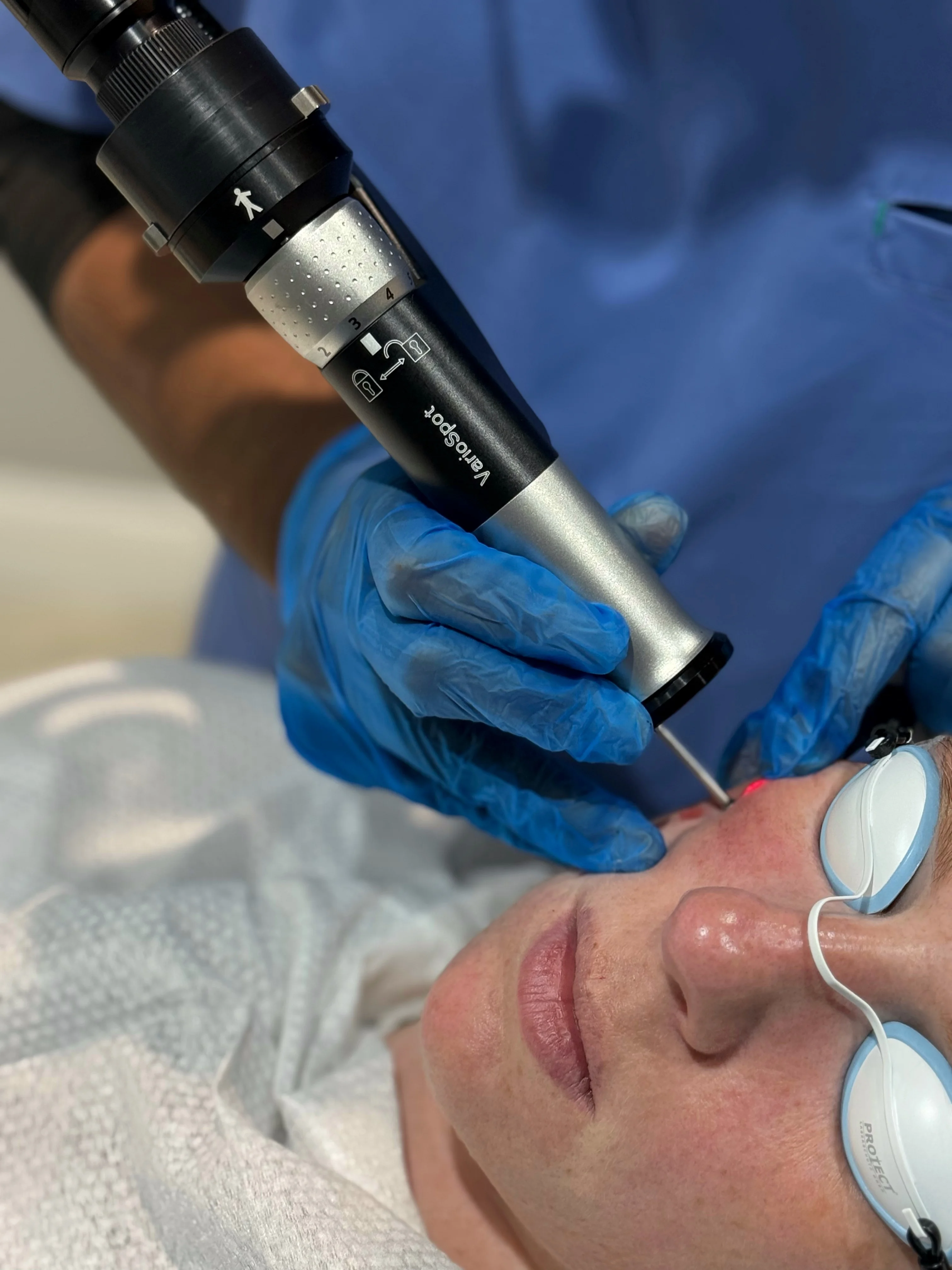
For an individual raised lesion, a precise laser skin lesion removal approach can remove the spot in a single, well-directed treatment. Dr Nick selects the most appropriate method for the size, depth and site of your lesion — the goal is clean removal with the lowest reasonable risk of a mark. The exact device used is confirmed at your consultation once the lesion has been assessed.
- Best for Single or raised lesions
- Sessions Usually 1
- Downtime Small scab over days
- How it works Targeted removal of the spot
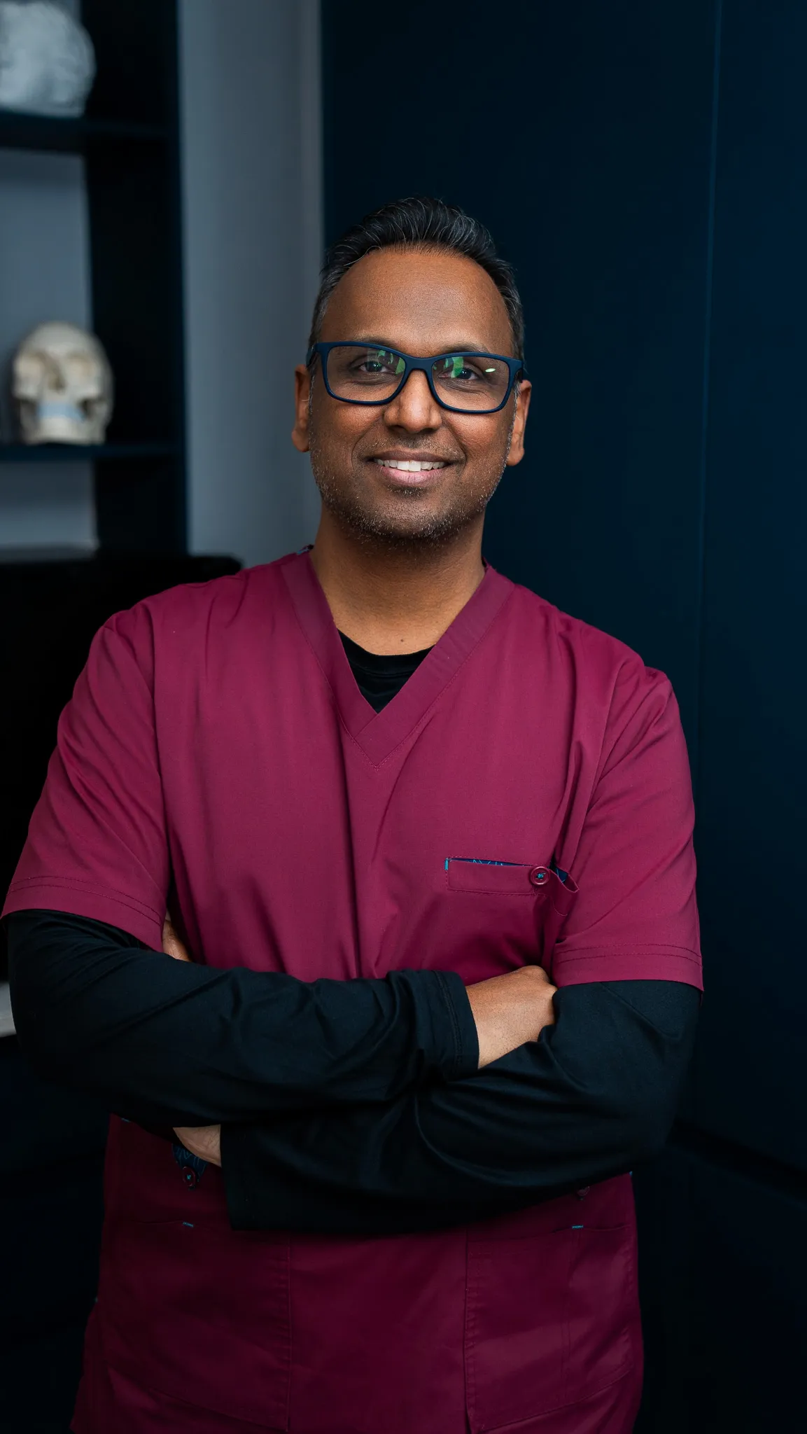
A red spot is exactly the kind of lesion that should be confirmed by a doctor before it is removed — we assess every one first, and refer appropriately if anything looks atypical.
Because the right method, the number of spots and the number of sessions vary from person to person, your price is confirmed at your consultation — once Dr Nick has assessed the lesion and agreed a plan with you. Your consultation covers the diagnosis (confirming the spot is benign), the recommended approach, realistic outcomes and a clear, honest quote with no surprises.
Why choose Rejuvena for cherry angioma removal in Bedford?
Assessed before treated
Dr Nick confirms every lesion is a benign cherry angioma before removal — the correct, doctor-led pathway that cryo-only chains do not advertise.
Doctor-led, GMC-registered
Dr Nick is MBBS, MRCGP, Diploma in Dermatology, GMC 6072251 — supported by our skin therapist Lana.
5★ from 161 reviews
Trusted by patients across Bedford, Kempston, Biddenham, Bromham, Clapham, Wootton, Great Denham and wider Bedfordshire.
Honest expectations
Realistic outcomes and clear aftercare — we remove the treated spot, and explain honestly that new spots can form over time.
Related treatments & concerns at Rejuvena Bedford
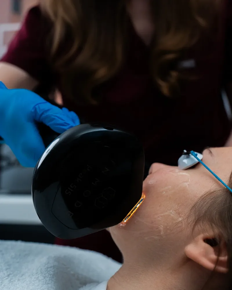
Lumecca IPL
Intense pulsed light for red and vascular concerns — a primary option for smaller and multiple cherry angiomas.
Learn more →Laser Skin Lesion Removal
Targeted removal of benign skin lesions, matched to the size and site of your spot.
Learn more →Thread Vein Removal
For linear or branching facial thread veins — a different vascular lesion to a cherry angioma.
Learn more →Venous Lake Removal
For the soft blue-purple lesions usually found on the lip or ear — another benign vascular concern.
Learn more →Verified Google reviews
What our patients say
5.0from 161 Google reviews
Lana and Dr Nick are both friendly and professional. I had treatment for thread vein removal and starting to see good results already! Thank you :)
Rejuvena repliedThank you very much for your kind words Alison. Your thread veins are improving great! Kind regards 🤗
Read this review on Google (opens in a new tab)I recently had a lesion removed from my cheek. The pigmented lesion had bothered me for years. I’m glad I finally decided to do something about it. I was quite nervous on the day, but Dr. Nick and Lana were so careful and reassuring — they really put me at ease. The procedure was quick and comfortable, and I feel so much better for having it done. I did a lot of research before I chose this clinic for my treatment. The clinic’s medical dermatology focus made me feel confident I would be in the right hands. I’m really grateful for the care I received and I would definitely recommend this clinic.
Rejuvena repliedThank you so much for your skin review Dupinder 🙏
Read this review on Google (opens in a new tab)I had lots of skin lesions on both hands and Dr Nick and Lana have been great treating me.I will definitely be returning for any future treatments and highly recommend seeing Dr Nick.Great service and facilities thanks again.
Rejuvena repliedAww! Thank you Gavin for your kind review. It was a great pleasure having you in our clinic and we look forward to seeing you in the future too. 😊🙏
Read this review on Google (opens in a new tab)Where to begin? Easy online booking, friendly team and professional pre-treatment consultations. Dr Nick’s medical background offers the expertise and reassurance that’s often missed in beauty salons. Lots of valuable advice as I explored thread vein treatments, options, budget, and desired results. In addition to the treatment itself, I’ve welcomed the superb after-care and support. I’ll absolutely be returning and already have my eye on their cool later facial. Thank you team Rejuvena!
Rejuvena repliedThank you very much Gemma, for your kind review. We are so glad to hear you liked our services. We are very grateful. ☺️🙏
Read this review on Google (opens in a new tab)I have been visiting Lana’s clinic for approximately one year, and each experience has been exceptional. I always leave feeling happy, confident, and fully satisfied with the quality of her work. Lana is highly skilled, attentive, and genuinely cares about her clients, ensuring that every detail is addressed with great care. I wholeheartedly recommend her services. If you are considering booking an appointment, do not hesitate—you will be in excellent hands!
Rejuvena repliedThank you very much for your sweetest review Roxana. It means a lot to me that you like our services ☺️🙏🤗🥺
Read this review on Google (opens in a new tab)Very very good! Clean. Can’t wait to start my journey here Non judgmental
Rejuvena repliedAww! Thank you very much Peter ☺️🙏 for leaving us a 5 star review. Really appreciate your kindness. We can’t wait to help you in getting your confidence back! 🤗
Read this review on Google (opens in a new tab)Every review above is published on Google. Tap any card to read it there.
Cherry angioma removal FAQs — Bedford
What is a cherry angioma (Campbell de Morgan spot)?
What causes cherry angiomas to appear?
Are cherry angiomas dangerous or a sign of cancer?
Can cherry angiomas be removed permanently — do they grow back?
How are cherry angiomas removed — laser, IPL or another method?
Does cherry angioma removal hurt, and is there downtime or scarring?
How much does cherry angioma removal cost in Bedford?
How many sessions will I need, and can several be removed at once?
Why should I see a doctor before having a red spot removed?
Book cherry angioma removal in Bedford
Have your red spot assessed by Dr Nick at Rejuvena Bedford — book a consultation online or call us.